Anatomy Of Back Of Neck : Second Layer Of Back And Neck Muscles 1866 Illustration Stock Image C040 5297 Science Photo Library / The back anatomy includes some of the most massive and functionally important muscles in the human body.
byAdmin-
0
Anatomy Of Back Of Neck : Second Layer Of Back And Neck Muscles 1866 Illustration Stock Image C040 5297 Science Photo Library / The back anatomy includes some of the most massive and functionally important muscles in the human body.. Still, many individuals pay far too little attention to them. Cervical (neck) thoracic (upper and middle back) lumbar (lower back) sacrum (tailbone). The human spine is composed of 4 sections of vertebrae. Jul 26, 2021 · the lateral neck muscles, also called the lateral vertebral muscles, are a group of muscles that pass obliquely along the lateral sides of the neck. The majority of these nerves control the functions of the upper extremities and allow you to feel your arms, shoulder, and back of your head.
Cervical (neck) thoracic (upper and middle back) lumbar (lower back) sacrum (tailbone). Understanding lower back anatomy is key to understanding the root of lower back and hip pain. Spinal anatomy is a remarkable combination of strong bones, flexible ligaments and tendons, large muscles and highly sensitive nerves. Still, many individuals pay far too little attention to them. It is designed to be incredibly strong, protecting the highly sensitive nerve roots, yet highly flexible, providing for mobility on many different planes.
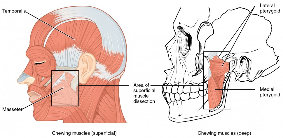
Axial Muscles Of The Head Neck And Back Anatomy And Physiology I from s3-us-west-2.amazonaws.com Cervical (neck) thoracic (upper and middle back) lumbar (lower back) sacrum (tailbone). The human spine is composed of 4 sections of vertebrae. It runs down the back part of the neck, and opens into the external jugular vein just below the middle of its course. The lumbar and sacrum region make up. The back anatomy includes some of the most massive and functionally important muscles in the human body. These include the anterior, middle and posterior scalene muscles , which extend between the transverse processes of the cervical vertebrae and the upper two ribs Understanding lower back anatomy is key to understanding the root of lower back and hip pain. Learn anatomy with free interactive flashcards.
Jul 23, 2016 · anatomy of back with organs, anatomy of organs from the back, anatomy of the back internal organs, anatomy of the lower back organs, anatomy of the organs in the back.
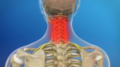
The human spine is composed of 4 sections of vertebrae. Still, many individuals pay far too little attention to them. The back anatomy includes some of the most massive and functionally important muscles in the human body. Cervical (neck) thoracic (upper and middle back) lumbar (lower back) sacrum (tailbone). The majority of these nerves control the functions of the upper extremities and allow you to feel your arms, shoulder, and back of your head. What are the lower back muscles and their anatomy? Jugularis anterior ) begins near the hyoid bone by the confluence of several superficial veins from the submaxillary region. May 06, 2015 · neck anatomy nerves picture there are 8 spinal nerves that originate from the cervical spine. Understanding the anatomy of your lower spine can help you communicate more effectively with the medical professionals who treat your lower back pain. Spinal anatomy is a remarkable combination of strong bones, flexible ligaments and tendons, large muscles and highly sensitive nerves. It is designed to be incredibly strong, protecting the highly sensitive nerve roots, yet highly flexible, providing for mobility on many different planes. It runs down the back part of the neck, and opens into the external jugular vein just below the middle of its course. Understanding lower back anatomy is key to understanding the root of lower back and hip pain.
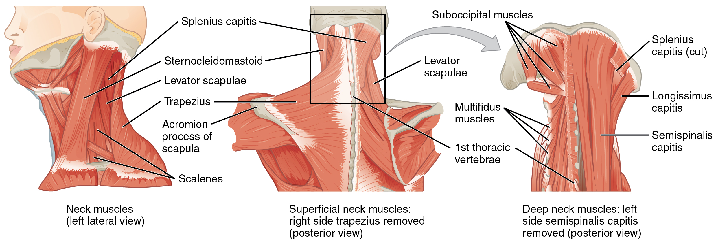
Apr 12, 2018 · the image below to shows all the major back muscles (as well as some neck muscles): The lumbar and sacrum region make up. May 06, 2015 · neck anatomy nerves picture there are 8 spinal nerves that originate from the cervical spine. The majority of these nerves control the functions of the upper extremities and allow you to feel your arms, shoulder, and back of your head. Understanding the anatomy of your lower spine can help you communicate more effectively with the medical professionals who treat your lower back pain.
Tips For Weekend Warriors Preventing Back And Neck Injuries Baldwin Bone Joint Pc from baldwinboneandjoint.com The majority of these nerves control the functions of the upper extremities and allow you to feel your arms, shoulder, and back of your head. Apr 12, 2018 · the image below to shows all the major back muscles (as well as some neck muscles): Spinal anatomy is a remarkable combination of strong bones, flexible ligaments and tendons, large muscles and highly sensitive nerves. May 06, 2015 · neck anatomy nerves picture there are 8 spinal nerves that originate from the cervical spine. It runs down the back part of the neck, and opens into the external jugular vein just below the middle of its course. It is designed to be incredibly strong, protecting the highly sensitive nerve roots, yet highly flexible, providing for mobility on many different planes. Learn anatomy with free interactive flashcards. Some important structures contained in or passing through the neck include the seven cervical vertebrae and enclosed spinal cord, the jugular veins and carotid arteries, part of the esophagus, the larynx
The anterior jugular vein ( v.
Understanding lower back anatomy is key to understanding the root of lower back and hip pain. Cervical (neck) thoracic (upper and middle back) lumbar (lower back) sacrum (tailbone). May 06, 2015 · neck anatomy nerves picture there are 8 spinal nerves that originate from the cervical spine. The lumbar and sacrum region make up. The majority of these nerves control the functions of the upper extremities and allow you to feel your arms, shoulder, and back of your head. Some important structures contained in or passing through the neck include the seven cervical vertebrae and enclosed spinal cord, the jugular veins and carotid arteries, part of the esophagus, the larynx The human spine is composed of 4 sections of vertebrae. These include the anterior, middle and posterior scalene muscles , which extend between the transverse processes of the cervical vertebrae and the upper two ribs It is designed to be incredibly strong, protecting the highly sensitive nerve roots, yet highly flexible, providing for mobility on many different planes. The back anatomy includes some of the most massive and functionally important muscles in the human body. Understanding the anatomy of your lower spine can help you communicate more effectively with the medical professionals who treat your lower back pain. The lumbar region of the spine, more commonly known as the lower back, is situated between the thoracic, or chest, region of the spine, and the sacrum. What are the lower back muscles and their anatomy?
Some important structures contained in or passing through the neck include the seven cervical vertebrae and enclosed spinal cord, the jugular veins and carotid arteries, part of the esophagus, the larynx The back anatomy includes some of the most massive and functionally important muscles in the human body. Spinal anatomy is a remarkable combination of strong bones, flexible ligaments and tendons, large muscles and highly sensitive nerves. Choose from 500 different sets of anatomy flashcards on quizlet. Understanding lower back anatomy is key to understanding the root of lower back and hip pain.
The Ventral Neck Muscles Lecturio Online Medical Library from d3uigcfkiiww0g.cloudfront.net Still, many individuals pay far too little attention to them. The lumbar region of the spine, more commonly known as the lower back, is situated between the thoracic, or chest, region of the spine, and the sacrum. What are the lower back muscles and their anatomy? Choose from 500 different sets of anatomy flashcards on quizlet. The human spine is composed of 4 sections of vertebrae. The lumbar and sacrum region make up. The back anatomy includes some of the most massive and functionally important muscles in the human body. It is designed to be incredibly strong, protecting the highly sensitive nerve roots, yet highly flexible, providing for mobility on many different planes.
May 06, 2015 · neck anatomy nerves picture there are 8 spinal nerves that originate from the cervical spine. Still, many individuals pay far too little attention to them. Learn anatomy with free interactive flashcards. The majority of these nerves control the functions of the upper extremities and allow you to feel your arms, shoulder, and back of your head. The lumbar region of the spine, more commonly known as the lower back, is situated between the thoracic, or chest, region of the spine, and the sacrum. The back anatomy includes some of the most massive and functionally important muscles in the human body. Some important structures contained in or passing through the neck include the seven cervical vertebrae and enclosed spinal cord, the jugular veins and carotid arteries, part of the esophagus, the larynx Jul 26, 2021 · the lateral neck muscles, also called the lateral vertebral muscles, are a group of muscles that pass obliquely along the lateral sides of the neck. The human spine is composed of 4 sections of vertebrae. Apr 12, 2018 · the image below to shows all the major back muscles (as well as some neck muscles): Understanding the anatomy of your lower spine can help you communicate more effectively with the medical professionals who treat your lower back pain. Spinal anatomy is a remarkable combination of strong bones, flexible ligaments and tendons, large muscles and highly sensitive nerves. It runs down the back part of the neck, and opens into the external jugular vein just below the middle of its course.